

What Your Lower Back Pain May Be Telling You About Your ure
Introduction
Lower back pain is among the most prevalent musculoskeletal complaints worldwide, affecting people of all ages and backgrounds. According to the World Health Organisation (WHO), musculoskeletal conditions, including lower back pain, are the leading contributors to disability globally. Actually, studies estimate that up to 80% of adults experience lower back pain at some point in their lives (Centres for Disease Control and Prevention (CDC)). Understanding what your lower back pain may be telling you about your ure is essential, as ure-related dysfunctions not only cause discomfort but can also signal underlying health issues with long-term implications.
Modern lifestyles, characterised by prolonged sitting, infrequent physical activity, and pervasive use of digital devices, have created an epidemic of ural misalignment. Below, we will explore the clinical connection between ure and lower back pain, clarify why these symptoms matter medically, and offer evidence-based advice for diagnosis, prevention, and management.
Overview and Definition
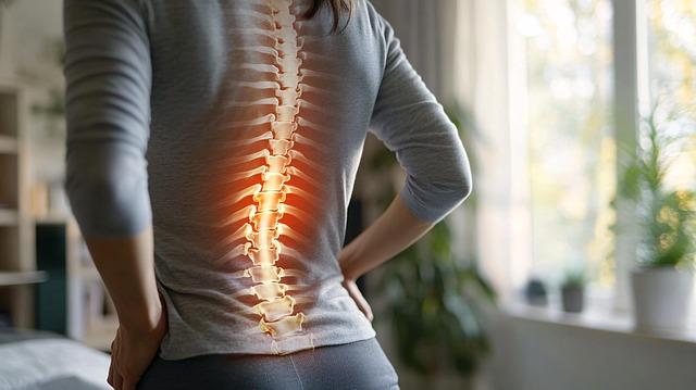
Lower back pain-clinically termed “lumbago”-is defined as pain, muscle tension, or stiffness localised below the costal margin and above the inferior gluteal folds, with or without leg pain (National Institutes of Health (NIH)). The lumbar spine, sacrum, associated muscles, ligaments, and neural elements are the primary anatomical structures involved. ure, in a biomechanical sense, refers to the alignment of the body’s joints and segments, especially how the pelvis and spine are stacked in relation to one another while standing, sitting, or moving.
According to systematic reviews published in The Lancet, lower back pain is the single leading cause of disability in 160 countries. Its lifetime prevalence ranges from 60% to 70% in industrialised nations, with a higher incidence among women and older adults. More than just a physical ailment, lower back pain affects overall well-being, limiting mobility, reducing productivity, and compromising mental health. Notably, chronic lower back pain is a risk factor for depression, sleep disturbances, and reduced quality of life (PubMed).
Causes and Risk Factors
The causes of lower back pain are multifactorial and often interwoven with individual ure. Below, we detail biological, genetic, environmental, and behavioural contributors, emphasising mechanisms that link ural habits with lower back dysfunction.
Biomechanical Factors and Poor ure
- Muscle Imbalances and Weakness: Prolonged poor ure-such as slouching or excessive lumbar lordosis, can inhibit core stabilisers (transversus abdominis, multifidus) and overload compensating structures (Harvard Health Publishing).
- Spinal Alignment Abnormalities: Kyphosis, lordosis, and scoliosis not only alter ure but disrupt normal vertebral loading, predisposing to disc degeneration and facet joint strain (Mayo Clinic).
- Prolonged Sedentarism: Office work, screen time, and driving promote static, flexed postures that weaken spinal extensors and compress lumbar discs (NIH Research).
Genetic and Structural Factors
- Intervertebral Disc Degeneration: Genetic predispositions influence disc hydration and elasticity, increasing susceptibility to injury with poor ure (PubMed).
- Congenital Anomalies: Vertebral malformations, such as spondylolysis or transitional vertebrae, may exacerbate ural imbalances and pain (MedlinePlus).
Environmental and Occupational Risks
- Ergonomics: Non-supportive chairs, high work surfaces, and other suboptimal workplace designs directly influence risk for both acute and chronic lower back pain (OSHA Ergonomics).
- Recurrent Lifting or Awkward Movements: Manual labour, repetitive twisting, or improper lifting techniques can accelerate degenerative changes and muscle fatigue (Healthline).
Comorbid and Psychosocial Factors
- Obesity and Metabolic Syndrome: Excess weight increases spinal load and predicts lower back pain, particularly with poor ural support (JAMA Network).
- Anxiety and Depression: Mental health conditions can heighten pain perception, promote muscle tension, and disrupt body awareness, indirectly worsening ure (Mayo Clinic FAQ).
Pathophysiology: How ure Affects the Lower Back
the spine’s natural curves-cervical lordosis, thoracic kyphosis, and lumbar lordosis-are designed to distribute body weight evenly and absorb shocks. Poor ure disrupts this equilibrium, directly affecting musculoskeletal structure and function:
- Increased Intervertebral Disc Pressure: Slouching or prolonged flexed sitting positions raise lumbar disc pressures, accelerating degeneration and risk for herniation (NIH Review).
- Muscle Fatigue and Overuse: Asymmetric muscle activation (e.g., tight hip flexors, weak gluteals) causes chronic fatigue and trigger points in the lower back (Mayo Clinic Treatment).
- Ligamentous Strain: Overextension or continuous flexion stretches spinal ligaments, causing microtrauma and pain sensitization.
- Nerve Impingement: Severe ural distortions may narrow the spinal or intervertebral foramina, compressing nerve roots, and causing radiculopathy (e.g., sciatica).
Scientific research has confirmed that upright,neutral spine ure decreases mechanical stress on discs and joints,supporting pain relief and prevention (Harvard Health Blog).
Clinical Presentation: Signs and Symptoms
Lower back pain associated with poor ure can manifest in several ways, from intermittent aches to disabling chronic discomfort. Key symptomatology includes:
- Dull, aching pain localized to the lumbar region or radiating to the buttocks and upper thighs.
- Stiffness, especially upon waking or after prolonged sitting.
- Sharp, shooting pain during specific movements, especially bending or twisting.
- Fatigue in the lower back muscles.
- Pain relief with ural correction, stretching, or walking.
In certain specific cases, ural lower back pain might potentially be accompanied by numbness, tingling, or weakness if nerve involvement occurs. Red flag symptoms-such as bowel or bladder incontinence, unexplained weight loss, or night sweats-may indicate a more serious pathology and warrant immediate medical assessment (NHS).
Diagnosis: Evaluating ural Causes of Lower Back Pain
proper diagnosis is essential to distinguish ure-related pain from other causes such as fractures, malignancy, or infection.Evaluation includes:
- Medical History: Assessment of symptom onset, duration, triggers (e.g., work ure), and alleviating factors.
- Physical Examination: ural analysis (visual inspection, range of motion, palpation for tenderness, muscle strength testing).
- functional Movement Assessment: Evaluates gait, squat mechanics, and core stability.
- Imaging Studies: X-rays or MRI might potentially be indicated if neurological symptoms or structural abnormalities are suspected (Mayo Clinic Diagnosis).
Standardized tools such as the Oswestry Disability Index and roland-Morris Disability Questionnaire can objectively gauge pain severity and functional impairment (NIH outcome Measures).
ural Patterns Frequently Associated with Lower Back Pain
Several common ural profiles predispose individuals to chronic lower back pain:
| ural type | Description | Associated Lower Back Issues |
|---|---|---|
| Hyperlordosis (“Swayback”) | Exaggerated inward lumbar curve,anterior pelvic tilt | facet joint compression,disc degeneration |
| Flat Back | Loss of natural lumbar curve,erior pelvic tilt | Disc herniation risk,muscle fatigue |
| Kyphosis with Forward Head | Rounded upper back,forward head shift | thoracolumbar muscle strain,compensatory pain |
| scoliosis | Abnormal lateral spinal curvature | Asymmetric muscle loading,chronic pain |
Prevention: How to Improve ure and Reduce Lower Back Pain Risk
evidence-based prevention strategies focus on ergonomic interventions,physical activity,and the cultivation of ural awareness.Key recommendations include:
- Ergonomic Optimization: Adjust workstation height/chair, use lumbar support, and place screens at eye level (OSHA Ergonomics).
- Movement Breaks: Stand, stretch, or walk for at least five minutes every hour during prolonged sitting (Medical News Today).
- Exercise Therapy: Core strengthening, Pilates, yoga, and functional movement retraining improve trunk stability and spinal alignment (Harvard Health Exercise).
- Weight Management: Maintaining healthy body weight reduces lumbar strain and supports musculoskeletal health.
- Footwear and Support: Wear supportive shoes, especially when standing or walking for long periods (Mayo Clinic on Prevention).
Management and Treatment Options
Acute and chronic lower back pain require individualized treatment, aligned with evidence-based guidelines (NIH Review):
Non-Pharmacologic Interventions
- Physical Therapy: Manual therapy,ural correction exercises,neuromuscular re-education (CDC Guidelines).
- Behavioral Therapy: Cognitive-behavioral therapy (CBT) for chronic pain management and enhancing self-efficacy (NIH CBT for Pain).
- Integrative Approaches: Acupuncture, mindfulness-based stress reduction, and gentle yoga.
Pharmacologic and Interventional Therapies
- Analgesics: NSAIDs and acetaminophen remain first-line for short-term symptom control (NHS on Painkillers).
- Muscle Relaxants and adjuncts: Prescribed for severe spasms or if sleep disturbed.
- Injections or Surgery: Reserved for refractory cases with nerve compression or structural instability.
It is indeed vital to avoid over-reliance on opioids or bed rest, as both are associated with inferior outcomes and higher risk of chronicity (CDC Guidelines).
When to see a Healthcare Provider
seek prompt medical assessment if your lower back pain:
- Persists beyond six weeks or worsens over time
- Is associated with neurological deficits (numbness, weakness, incontinence)
- Emerges after trauma, falls, or is accompanied by fever, unexplained weight loss
Diagnostic evaluation by a primary care provider, physical therapist, or orthopedic specialist ensures early identification of non-ural causes and initiates appropriate management (NHS: When to See a GP).
Prognosis and Outcomes
Most cases of ure-related lower back pain improve within weeks when managed with conservative therapy and ural correction. However, up to one-third of cases may develop chronic symptoms, particularly if ergonomic and lifestyle risk factors persist (Harvard Health).
Long-term prognosis improves with multidisciplinary care, physical rehabilitation, and proactive lifestyle modification. For severe or recurring cases, individualized treatment planning guided by healthcare professionals is essential (Mayo Clinic).
Conclusion
Your lower back pain can be an important health signal, especially when it reflects poor ure. Recognizing the biological and mechanical relationship between spinal alignment and musculoskeletal health empowers individuals and clinicians to act early-reducing pain, disability, and the risk of chronicity. Through ergonomic awareness, routine movement, targeted exercise, and timely medical consultation, most ure-related lower back disorders can be effectively prevented and managed.
For further information, refer to:
Frequently Asked Questions (FAQs)
Can bad ure really cause long-term back problems?
Yes, prolonged poor ure can lead to spinal misalignment, chronic muscle strain, and even degenerative disc disease over time (Healthline).
How do I tell if my lower back pain is due to ure?
If your pain worsens with sitting or standing in one position and improves with movement or stretching, ural factors are likely involved. A healthcare provider can help identify specific ural imbalances (Medical News Today).
Can ure correction devices help?
ure devices may offer temporary support, but lasting improvement comes from strengthening core muscles, optimizing ergonomics, and practicing body awareness. Use devices as adjuncts, not substitutes for active intervention (harvard Health Blog).
Should I avoid exercise when I have lower back pain?
Gentle movement and low-impact exercise are generally beneficial and recommended unless contraindicated by your physician. Avoid high-impact or twisting activities until cleared by a professional (NHS).
When do I need imaging for lower back pain?
Imaging is indicated if pain persists beyond six weeks, is severe, or accompanied by neurological deficits. Discuss imaging with your healthcare provider to assess appropriateness (CDC).